


An eye and orbit ultrasound uses high-frequency sound waves to measure and produce detailed images of your eye and eye orbit (the socket in your skull that holds your eye).
An eye and orbit ultrasound, also known as an ophthalmologic ultrasound or echography, provides a much more detailed view of the inside of your eye than a routine eye exam.
An ultrasound technician or an ophthalmologist usually performs the procedure. An opthalmologist is a doctor who specializes in diagnosing and treating eye disorders and diseases.
You can receive this ultrasound in an office, outpatient imaging center, or hospital.
Keep reading to learn what eye conditions an eye and orbit ultrasound can identify, how the procedure is performed, and what to expect.
Your eye doctor may order an eye and orbit ultrasound if:
- you’re experiencing unexplained problems with your eyes, like changes in vision
- you’ve recently sustained an injury or trauma to the eye area
- you’ve had results on another eye exam that the doctor recommends investigating further
- you have unexplained headaches
This procedure helps identify issues with the eyes and diagnose eye diseases. Some of the issues the test can help identify can include:
- foreign substances in the eye
- detachment of the retina
- tumors or neoplasms involving the eye
An eye and orbit ultrasound can also be used to help diagnose or monitor:
- glaucoma, a progressive disease that can lead to vision loss
- cataracts, cloudy areas in the lens of your eye
- lens implants, plastic lenses implanted in the eye after the natural lens has been removed, usually due to cataracts
Your doctor can also use this procedure to measure the thickness and extent of a cancerous tumor and determine treatment options.
An eye and orbit ultrasound requires no specific preparation.
The procedure usually causes no pain. A healthcare professional usually gives you anesthetic drops to numb your eye and minimize discomfort.
While your pupils won’t be dilated, your vision may be temporarily blurred during the test. You should be able to drive 30 minutes after the ultrasound, although you may feel more comfortable arranging for someone else to drive.
Your eye doctor will advise you not to rub your eyes until the anesthetic has completely worn off. That’s to prevent you from unknowingly scratching your cornea.
There are two parts to an eye and orbit ultrasound. The A-scan ultrasound takes measurements of your eye. The B-scan allows the doctor to see the structures in the back of your eye.
The combined procedure (A and B scans) will take 15 to 30 minutes to complete.
A-scan
The A-scan measures the eye. This helps determine the correct lens implant for cataract surgery.
While sitting upright in a chair, a doctor will likely have you place your chin on a chin rest and look straight ahead. An oiled probe will be placed against the front portion of your eye as it’s scanned.
A healthcare professional can also perform an A-scan while you’re lying down. In that case, a fluid-filled cup, or water bath, is placed against the surface of your eye as it’s scanned.
B-scan
The B-scan helps your doctor see the space behind the eye and certain structures in the eye, such as the retina and vitreous. It can also let an opthalmologist see the back wall of the eye, or posterior sclera, and the optic nerve.
This scan is helpful if health conditions, including cataracts, make it difficult to see the back of the eye.
The B-scan also helps diagnose and stage tumors, retinal detachment, and other conditions.
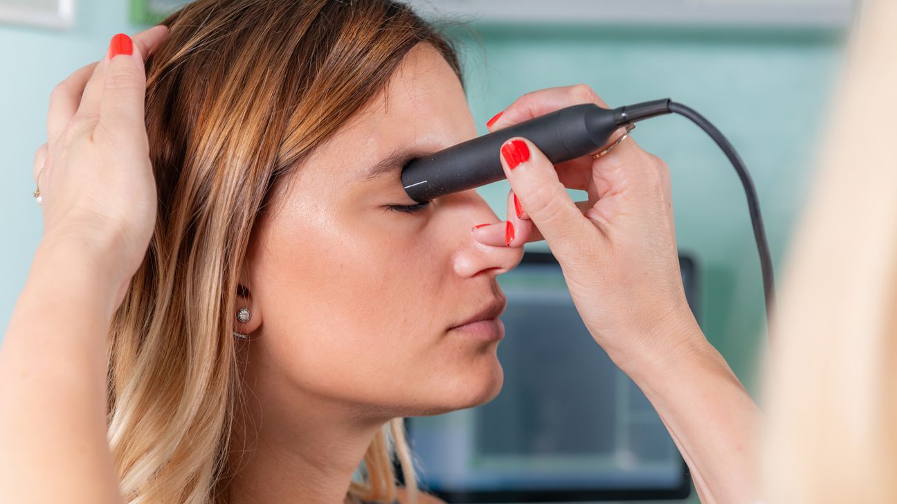
During a B-scan, you’ll usually sit with your eyes closed. The eye doctor then applies a gel to your eyelids. Your eye doctor then places the probe against your eyelids. They usually direct you to keep your eyes closed while you move your eyeballs in different directions.
An echography is typically a quick and painless procedure with no serious side effects or risks.
After they finish the ultrasound, the ophthalmologist will likely review the results with you.
Your doctor will determine if the eye measurements taken from the A-scan are within the normal range.
The B-scan gives your doctor structural information about your eye. If the results are abnormal, the doctor usually uses the results to determine the cause.
Some conditions that the B-scan might reveal include:
- foreign bodies in the eye
- cysts
- swelling
- detachment of the retina
- damaged tissue or injury to the eye socket (orbit)
- vitreous hemorrhage (bleeding into the clear gel, called the vitreous humor, that fills the back of the eye)
- cancer of the retina, under the retina, or in other parts of the eye
Once the doctor reaches a diagnosis, they will work to determine the best course of treatment for you.
An eye and orbit ultrasound is a detailed eye exam performed by an opthalmologist. It can detect certain health conditions affecting the eyes, including a detached retina or eye injury. Doctors may also use it to monitor other eye conditions, such as cataracts and glaucoma.
The ultrasound is usually quick and pain-free.