


Tomosynthesis is a new and advanced form of breast cancer screening. It can help detect early signs of breast cancer in people with no symptoms. It is most helpful for those with dense breasts.
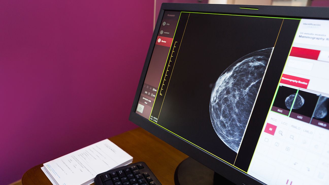
Tomosynthesis is an advanced type of mammography. During tomosynthesis, the machine takes multiple images of the breast, which are sent to a computer. The computer uses an algorithm to combine them into a 3D image of the entire breast.
Other names for tomosynthesis include:
- 3D mammography
- breast tomosynthesis
- digital breast tomosynthesis (DBT)
- tomo
Tomosynthesis and mammography are similar. They both are imaging techniques that doctors use to detect signs of breast cancer. Doctors use both techniques during annual exams and to check the progression of breast cancer.
But, healthcare professionals consider tomosynthesis a more advanced and detailed imaging technique than traditional mammography.
A traditional mammogram only captures a 2D image. In tomosynthesis, doctors are able to analyze
The 3D imaging in tomosynthesis helps doctors to see small lesions and other signs of breast cancer earlier than a traditional mammogram.
Other benefits of using tomosynthesis include the following:
- more accurate and less likely to result in false positives
- greater accuracy when screening for breast cancer in people with dense breasts
- early detection of breast cancer in people experiencing breast cancer symptoms
Tomosynthesis can help detect breast cancer years before people start to experience any symptoms or a doctor can see any signs.
Tomosynthesis is still a relatively new procedure, and not all imaging technicians or doctors will be familiar with it.
The possible risks of using tomosynthesis instead of traditional mammography may include:
- More radiation in some cases: Radiation levels will depend on the mammography machine. For some people receiving 3D mammograms, there may be slightly more exposure to radiation due to more images being taken of each breast. However, the radiation levels are still low enough to meet the Food and Drug Administration (FDA) safety standards. The radiation leaves your body shortly after the procedure.
- Inconsistent reconstruction algorithms: Specific algorithms for constructing the 3D images may vary, which may affect your results.
- Variation in the images: The arc of the movement of the X-ray tube may vary, which may cause variation in the images.
Insurance providers in the United States typically cover the cost of traditional mammography.
Many providers, including Medicare, now cover tomosynthesis as part of breast cancer screening.
If you have symptoms of breast cancer and will require a diagnostic exam instead of a screening, you may have to pay for some of your mammogram or tomosynthesis. This will depend on your insurance provider as well as your location.
If you don’t have insurance, breast cancer screening costs can vary, depending on the state you are in and your age.
A 2020 study looking at Blue Cross Blue Shield customers in their 40s found that initial breast cancer screening and follow-up tests cost an average of
A 2018 study used medical claims from 2011 to 2015 to estimate the cost of breast imaging and diagnostic procedures. The researchers found that, on average, a 2D diagnostic mammogram costs $354 for people ineligible for Medicare. Diagnostic tomosynthesis costs $136 for people ineligible for Medicare.
The insurance companies covered part of these costs, while the individuals covered the rest.
Learn more about the cost of mammograms — and where to find low cost or free ones.
Preparing for tomosynthesis is similar to preparing for a traditional mammogram. Some tips to follow are listed below.
Before you arrive:
- Request your prior mammograms. This allows the doctor to compare both images to better see any changes that may occur in your breasts.
- Schedule the procedure 1 to 2 weeks after your period starts to help reduce breast tenderness.
- Let the doctor and imaging technician know if you think you may be pregnant or if you’re nursing. The doctor may want to use a different procedure or take additional precautions.
- Let the doctor and imaging technician know about:
- any symptoms you may be having
- surgeries to or near your breasts
- a family history of breast cancer
- personal hormone use
- Let the doctor and imaging technician know if you have breast implants.
- Reduce the amount of caffeine you eat or drink prior to your procedure, or eliminate it entirely. This also helps reduce possible breast tenderness.
What to wear:
- Consider wearing 2-piece clothing. This makes undressing for the procedure easier and allows you to remain dressed from the waist down.
- Avoid using deodorant, powder, lotion, oil, or cream from the waist up on the day of the procedure.
The day of your procedure:
- Let the doctor and imaging technician know, once again, if you:
- are nursing or think you may be pregnant
- are having any symptoms
- have had surgeries to or near your breasts
- have a family history of breast cancer or a personal history of hormone use
- have breast implants
- Ask when you should expect the results
The procedure for tomosynthesis is very similar to that of traditional mammography.
Tomosynthesis even uses the same type of imaging machine as traditional mammography. However, the images it takes are different. Not all imaging machines are equipped to take tomosynthesis images.
Overall, the procedure can last up to 30 minutes.
Here’s what you should expect:
- When you arrive, the technician will take you to a changing room to remove your clothes from the waist up and provide you with a gown or cape.
- Then, they will take you to the same machine or type of machine that performs a traditional mammogram. The technician will position one breast at a time in the X-ray area.
- Your breasts will be tightly compressed like they are during a traditional 2D mammogram.
- They will position the X-ray tube over your breast.
- During the procedure, the X-ray tube will move by making an arc over your breast.
- During the procedure, they will take approximately 11 images of your breast in 7 seconds.
- You’ll then change positions so that images can be taken of your other breast.
- After the procedure is complete, your images will be sent to a computer that will make a 3D image of both breasts, also known as bilateral tomosynthesis.
- The final image will be sent to a radiologist to interpret the results.
After the procedure is complete, you can resume your usual activities and diet.
If your results show no signs of cancer, you may hear from the doctor that same day.
If your results suggest you may have cancer, further testing and follow-up are needed. Result times will vary by facility.
If the results are inconclusive, a doctor may call you back in for further tests, such as an MRI or ultrasound. The results of one of those tests may take at least 1 week to receive. However, according to a
Tomosynthesis is most helpful in screening for breast cancer in people with dense breasts.
If you have dense breasts or possible symptoms of breast cancer, consider discussing with a doctor the option of having tomosynthesis imaging done in addition to or instead of a traditional mammogram.
Tomosynthesis is still a relatively new procedure, so it’s not available at all facilities that perform mammography. Be sure to ask a doctor or imaging center if this option is available for you.